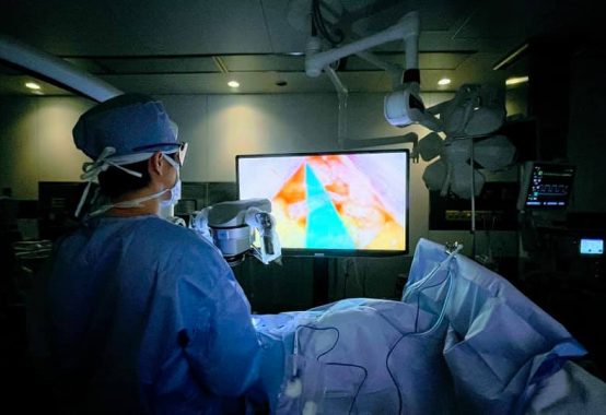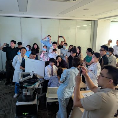
2019年より亀田リンパ浮腫センターで使用している、最新のOCT (光干渉断層撮影)顕微鏡を用いたLVAにおける手術中リンパ管選択・吻合部評価についての論文が、正式にpublishされました!
OCTを用いたマイクロサージャリーに関して世界初の英語論文になります。
約2年前から少しずつ積み重ねてきた研究ですが、沢山の方々の支えがあり、ここまで来れました。
OCTは、マイクサージャリーやリンパ浮腫手術のレベルを一段と引き上げる、変革をもたらすものと確信しています。
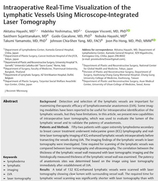
Our latest research paper, “Intraoperative Real-Time Visualization of the Lymphatic Vessels Using Microscope-Integrated Laser Tomography”, has been published in the Journal of Reconstructive Microsurgery!
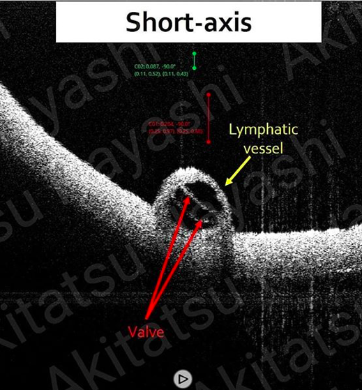
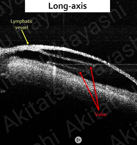
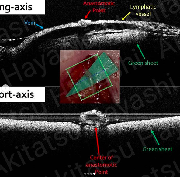
It is the first report that used Laser Tomography (OCT) to evaluate real-time not only lymphatic degeneration status but also patency of the LVA sites intraoperatively for maximizing the effect of LVA.
We strongly believe this advanced imaging technology is going to evoke a paradigm shift in the field of supermicrosurgery.
-My three must-have tools for the most effective LE surgery-
1. ICG lymphography (Pre-Op)
2. Ultra High- frequency Ultrasound (Pre-Op)
3. ★ Laser Tomography (Intra-Op)★