Latest Diagnostic Imaging
In 2017, we established and presented the world’s first method of using ultra-high frequency ultrasound to diagnose lymphatic vessels and perform surgeries more effectively and efficiently. This technology has made it possible to provide treatment without the use of contrast media during surgery, greatly reducing burden on the body.
Evaluating Lymphatic Vessels and Lymphedema Using Ultra-High Frequency Ultrasound
Lymphedema progresses with the degeneration of lymph vessels. For more details, please refer to the separate page Progression and Degeneration of Lymphedema.

In this section, we will compare the diagnosis of lymphedema using conventional high-frequency ultrasound and a new diagnosis method called ultra-high frequency ultrasound.
Diagnosis of Lymphedema Using Conventional High-Frequency Ultrasound
「The conventional evaluation method using high-frequency ultrasound is effective in the routine treatment of lymphedema, as it can determine the severity of lymphedema and the timing of its deterioration by regularly observing the condition of the skin, subcutaneous tissue, and fluid retention.
However, on the other hand,
- It is difficult to detect lymphatic vessels that are not dilated.
- It is difficult to distinguish φ0.3-0.4 mm or less from other tissues.
- It is not possible to evaluate the quality of lymphatic vessels.
The following are the phases that can be identified by high-frequency ultrasound:

Range of identification by high-frequency ultrasound
Diagnosis of lymphedema using ultra-high frequency ultrasound
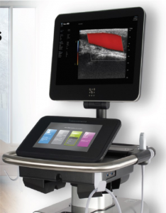
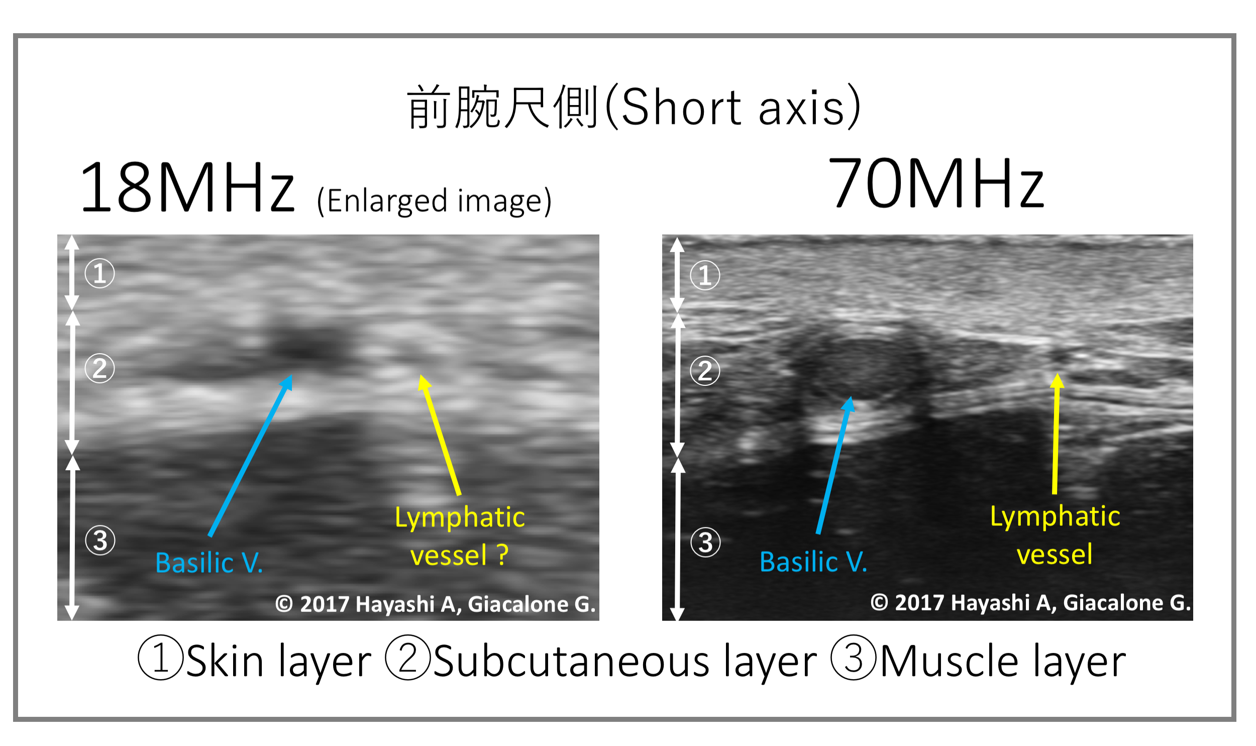
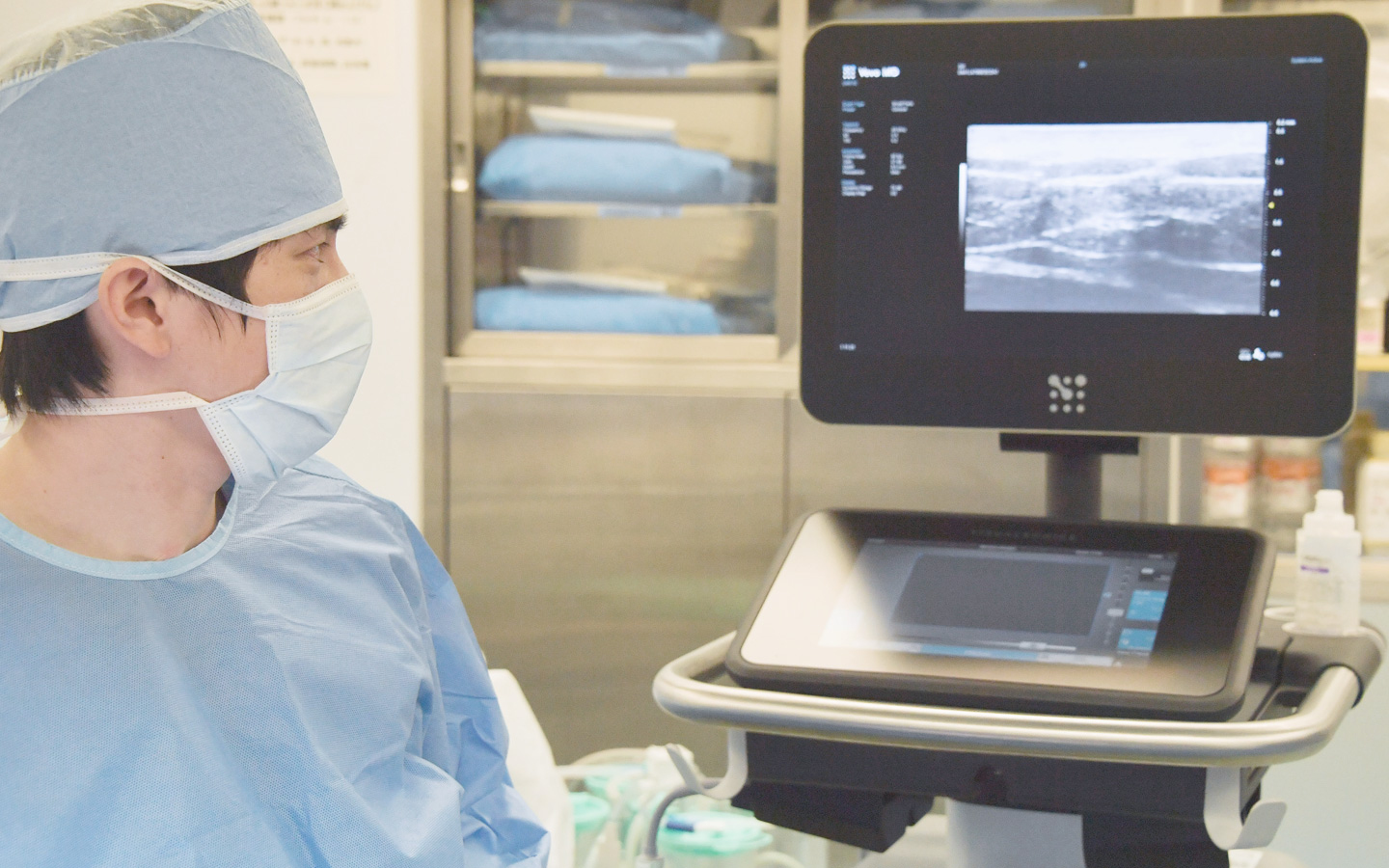
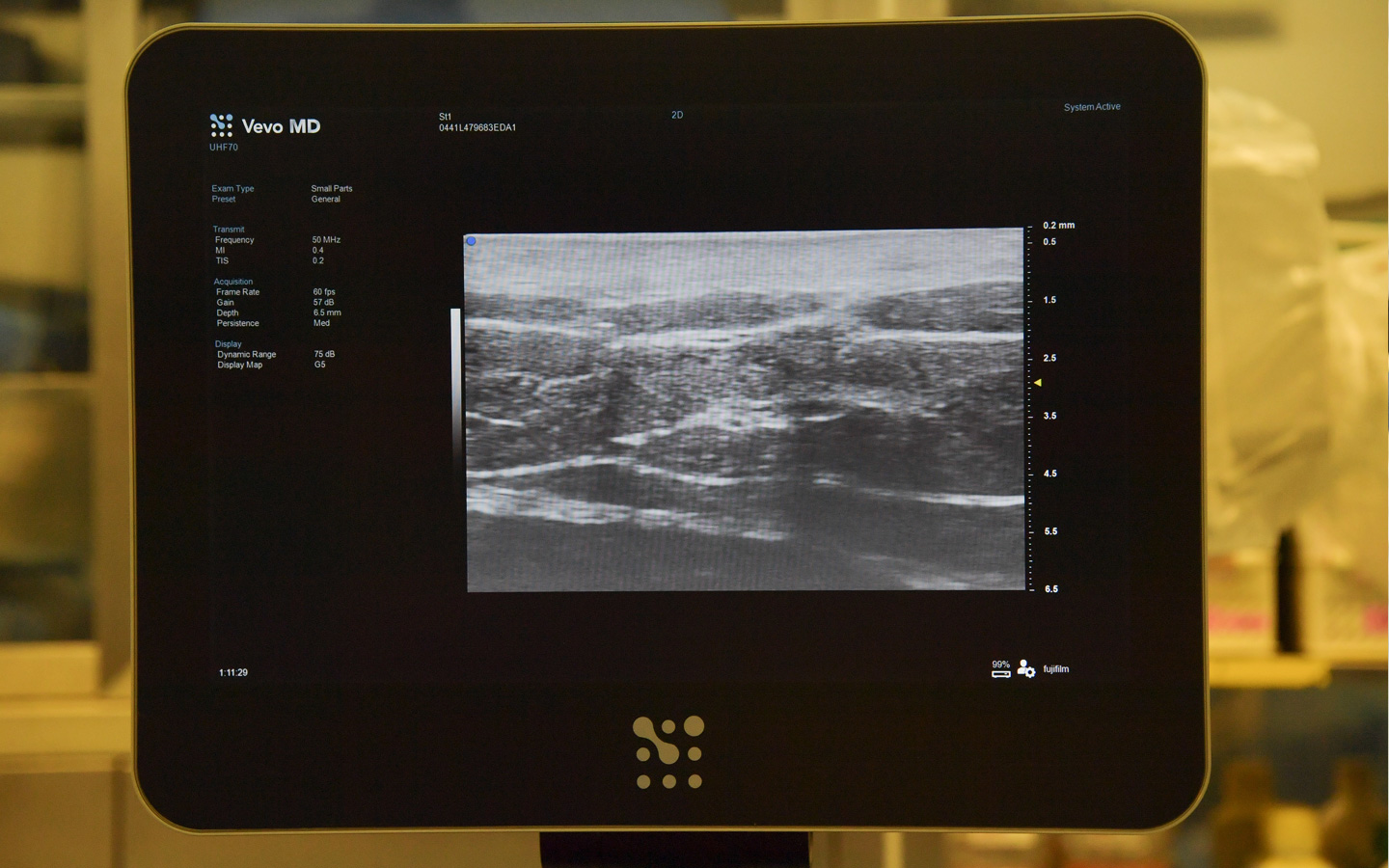
In the latest lymphedema evaluation method using-ultra high-frequency ultrasound, ultra-high frequency scans of up to 70 MHz can be performed in real time with high resolution. Furthermore, we are now able to capture the degeneration of lymph vessels, which was difficult to do with conventional high-frequency ultrasound, and to identify lymph vessels in a wider phase of lymphedema progression.

Identification range via ultra-high frequency ultrasound
Ulnar side of the forearm
Minimally invasive and efficient/effective lymphedema diagnosis and surgery are now possible
he use of ultra-high frequency ultrasound has made it possible to capture the changes in lymphatic vessels, and is expected to be used for non-invasive and efficient diagnosis and treatment as well as for the development of new surgical methods.
関連記事
むくみとリンパ浮腫
原因・発症・進行と症状
診断
治療法
術後
手術の注意点・合併症
予防と運動療法

Akitatsu Hayashi
Kameda Medical Center / Kameda Kyobashi Clinic
Director of Lymphedema Center
Dr. Hayashi presented state-of-the-art lymphatic vessel imaging technology using Ultrasound and Optical
Coherence Tomography (OCT) for the first time in the world. He focuses on the spread of more efficient and
effective lymphedema surgery using the technology.Check profile more


